Fraunhofer Institute for High-Speed Dynamics, Ernst-Mach-Institut, EMI
Medical technology
A Less Debilitating Approach to Cancer Diagnosis
Less radiation exposure during diagnosis and treatment for breast and lung cancer: New Fraunhofer method combines X-ray imaging and radar.
Imaging methods are an essential part of medicine, used in everything from diagnosis and treatment to follow-up examinations. X-ray mammography dominates the field of early detection of breast cancer, providing rapid and accurate two-dimensional images. Three-dimensional computed tomography (CT) is used in cancer diagnosis, but because it involves high doses of radiation, it poses health risks of its own. While a person’s nat-ural annual radiation exposure is about 2.1 millisieverts, a chest CT involves about three times as much. In the MultiMed project, which explores multimodal medical imaging in 3D, Fraunhofer researchers are developing a method that combines X-ray imaging and radar. It is not only expected to improve the accuracy and efficacy of diagnosing, moni-toring and treating breast and lung cancer but also lighten the burden on patients
enhances the quality and level of detail of the CT images, reduces disruptive artifacts and lowers radiation exposure.
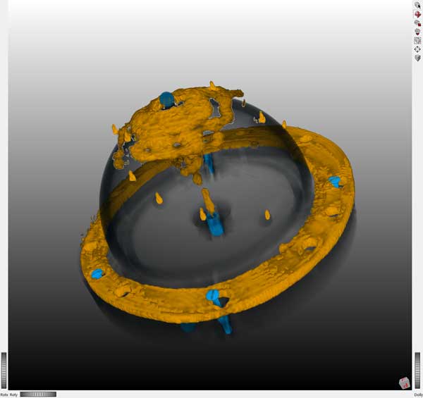
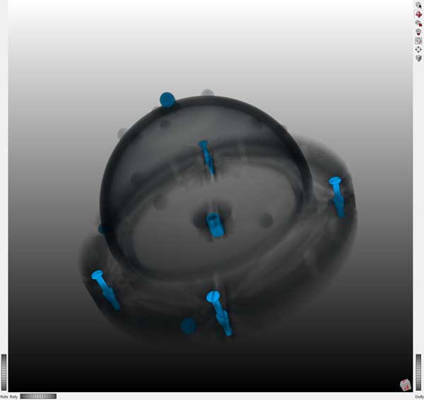
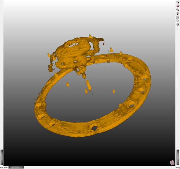
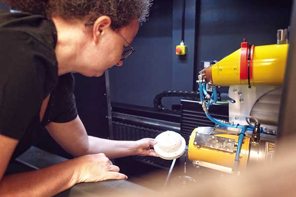
The research team has already developed initial imaging phantoms to test this method. Phantoms like these are artificial models that realistically simulate tissue structures, sup-plying suitable signals for radar and X-ray measurements.
Radar in medicine: an outsider with potential
Radar is established in many areas: Airports use it to monitor air traffic, and radar sen-sors are a part of automotive assistance systems. In medicine, however, this method has been more of an outsider so far. And yet, it too can supply three-dimensional im-ages — without posing any health risks. Radar does have lower resolution, plus it can-not penetrate as deep when compared to other methods. But it provides material infor-mation that other processes cannot offer directly. Radar detects differences in electrical permittivity and conductivity, so it can be used to identify changes in tissue.
The challenge lies in combining the types of measurements taken by the two methods. The researchers are developing special ways of linking the imaging data from the two systems together. Called “co-registering,” this approach sets the radar and X-ray data in spatial relation to each other.
To further enhance image quality and visualize interior areas of the body in three di-mensions via radar, the researchers are working on new radar reconstruction algo-rithms. This will unlock sharper images and more detailed insight into tissue properties.
At the same time, X-ray CT reconstruction is also being optimized: Radar data is being incorporated into the X-ray reconstruction to produce a multimodal CT algorithm. This enhances the quality and level of detail of the CT images, reduces disruptive artifacts and lowers radiation exposure.
The research team has already developed initial imaging phantoms to test this method. Phantoms like these are artificial models that realistically simulate tissue structures, sup-plying suitable signals for radar and X-ray measurements.
Goal: detect tissue changes early on, accurately and with low radiation
The three-year project is intended to result in a multimodal lab system. This testing and development environment will combine CT imaging using X-rays with radar imaging to provide more comprehensive and accurate analyses. “The new approach has the po-tential to detect tissue changes early on and with great accuracy — and with much less of a burden on patients than before,” says project manager Victoria Heusinger-Hess from the Fraunhofer Institute for High-Speed Dynamics, Ernst-Mach-Institut, EMI.
The Fraunhofer-Gesellschaft is funding the three-year research project. Under the lead-ership of Fraunhofer EMI, the Fraunhofer Institute for Digital Medicine MEVIS and the Fraunhofer Institute for High Frequency Physics and Radar Techniques FHR are also in-volved.